Upper Leg Tendon Anatomy : The muscles located within the posterior compartment of the thigh are the biceps femoris, semitendinosus and semimembranosus.
byAdmin-
0
Upper Leg Tendon Anatomy : The muscles located within the posterior compartment of the thigh are the biceps femoris, semitendinosus and semimembranosus.. Upper leg muscle pain is a very hard pain affect the leg pain as a whole. The knee joint is commonly injured, so understanding its anatomy can help you understand the conditions that cause problems, so you stay safe and prepared. The hamstring muscles in the back of the thigh, the quadriceps muscles in the front, and the adductor muscles on the inside. •medial thigh muscles•adductor longus muscle•adductor magnus muscle•adductor. Upper leg tendon anatomy :
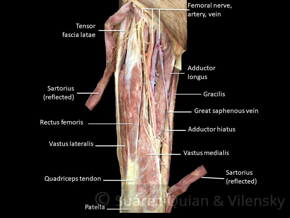
The vastus lateralis is a muscle located on the lateral, or outside, part of your thigh. When we knowing the causes of the upper leg muscle pain it will be simple to treat and relieve the pain. Upper leg tendon anatomy : Meanwhile, the vastus lateralis is on the side of the thigh, while the vastus intermedius is hidden below the rectus femoris(5). 430) is the most superficial muscle on the medial side of the thigh.
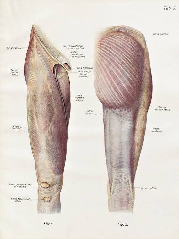
Muscle Compartments Of The Thigh Complete Anatomy from 3d4medical-cdn.s3-us-west-1.amazonaws.com The hamstrings are three muscles at the back of the thigh that affect hip and knee movement. The rectus femoris is located in the center of the thigh, while the vastus medialis is in the middle of the said body part. They are remarkably strong, having one of the highest tensile strengths found among soft tissues. It's the area that runs from the hip to the knee in each leg. Tendons are thick bands of tissue that connect muscles to bone. Tendons are also bands of connective tissue. The human leg, in the general word sense, is the entire lower limb of the human body, including the foot, thigh and even the hip or gluteal region. The vastus lateralis is a muscle located on the lateral, or outside, part of your thigh.
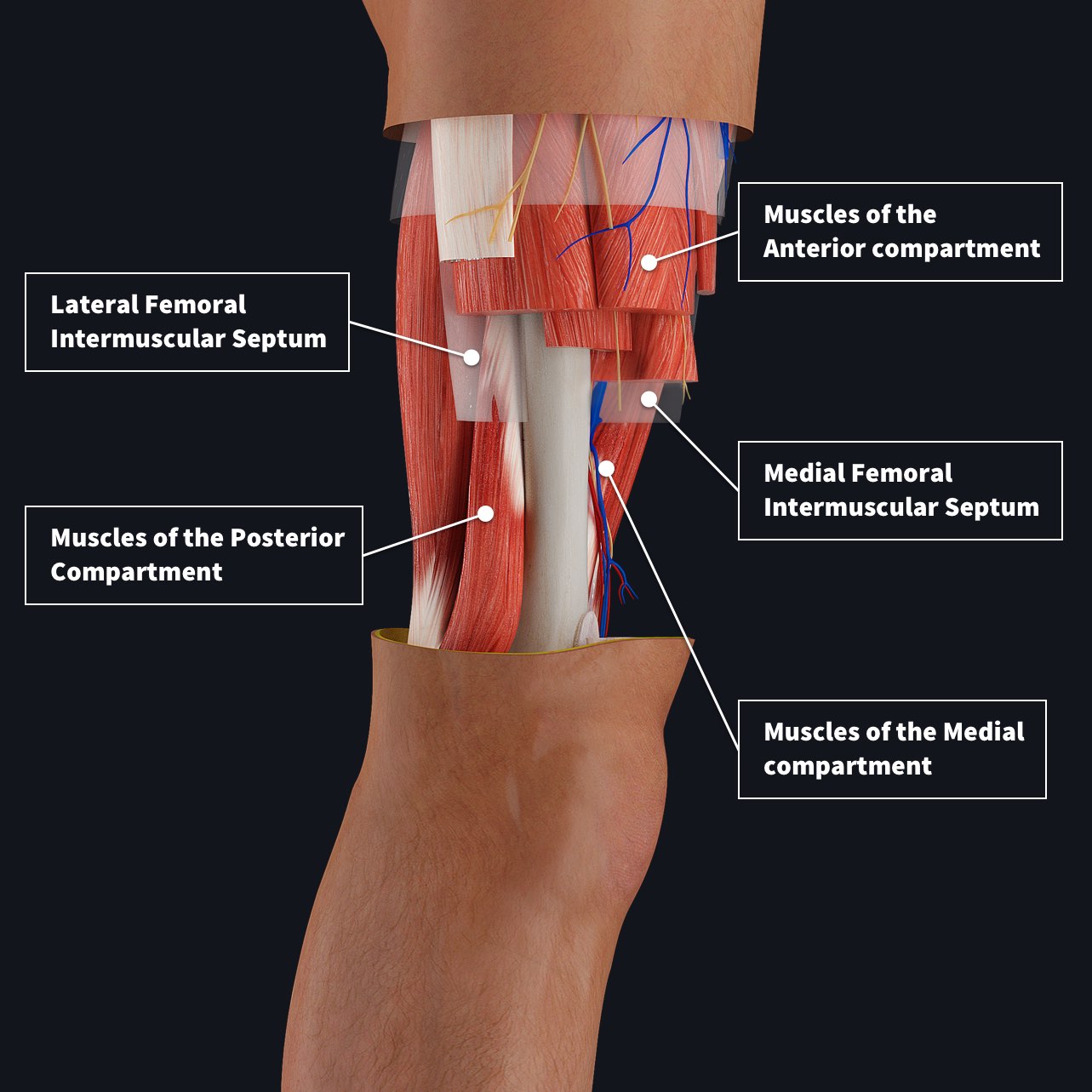
The hamstrings refer to 3 long posterior leg muscles, the biceps femoris, semitendinosus, and semimembranosus.
The largest muscle masses in the leg are present in the thigh and the calf. The hamstrings refer to 3 long posterior leg muscles, the biceps femoris, semitendinosus, and semimembranosus. The human leg, in the general word sense, is the entire lower limb of the human body, including the foot, thigh and even the hip or gluteal region. .upper leg muscles anatomy, upper leg muscle numbness, upper leg muscle pain weakness, upper leg muscle stiffness, upper leg muscles complex muscle and joint interaction innervation of the shoulder complex muscles of the shoulder. It's the area that runs from the hip to the knee in each leg. The hamstring portion of the adductor magnus has a similar action to these muscles, but is located in the medial thigh. The hamstring muscles in the back of the thigh, the quadriceps muscles in the front, and the adductor muscles on the inside. The four muscles all extend the lower leg. This is why you have to indicate which biceps you are taking about when discussing one or other of these muscles. •medial thigh muscles•adductor longus muscle•adductor magnus muscle•adductor. Upper leg anatomy and function. These muscles start at the bottom of your pelvis extending down the back of your thigh and along either side of your knee, to your lower leg bones. The iliopsoas muscle flexes your hip, bends your trunk towards your thigh and rotates your thigh bone.
It consists of several parts: Parts of the thigh your thigh is the area of your upper leg between your hip joint and your knee. On the medial edge of the posterior thigh is the gracilis muscle. Each of these muscles is a discrete organ constructed of skeletal muscle tissue, blood vessels, tendons, and nerves. The four muscles all extend the lower leg.
Upper Leg Muscles Ligaments C 1900 Antique Anatomy Print Etsy from i.etsystatic.com 3d anatomy tutorial on the muscles of the thigh and the gluteal region from anatomyzone for more videos, 3d models and notes visit: Your quadriceps muscles are in the front of your thigh. It's the area that runs from the hip to the knee in each leg. Lateral (fibular) collateral ligament (fcl) upper part middle part lower part popliteus tendon (pt) upper part i. Upper leg muscle pain is a very hard pain affect the leg pain as a whole. The hamstring muscles in the back of the thigh, the quadriceps muscles in the front, and the adductor muscles on the inside. The large achilles tendon is the most important tendon for walking, running we created an anatomical atlas of the upper limb, an. These muscles start at the bottom of your pelvis extending down the back of your thigh and along either side of your knee, to your lower leg bones.
The fibers run vertically downward, and end in a rounded tendon, which passes behind the medial condyle.
The human leg, in the general word sense, is the entire lower limb of the human body, including the foot, thigh and even the hip or gluteal region. The four muscles all extend the lower leg. Tendons are also bands of connective tissue. The largest muscle masses in the leg are present in the thigh and the calf. It is thin and flattened, broad above, narrow and tapering below. This is why you have to indicate which biceps you are taking about when discussing one or other of these muscles. This tendon helps your leg bend when you raise your knee. The hamstring portion of the adductor magnus has a similar action to these muscles, but is located in the medial thigh. Squeeze your knees together and boom, you're contracting the adductors. Tendons are thick bands of tissue that connect muscles to bone. Tendons are cords made of tough tissue, and they work as special connector pieces between bone and muscle. The muscles located within the posterior compartment of the thigh are the biceps femoris, semitendinosus and semimembranosus. It consists of several parts:
Tendons are cords made of tough tissue, and they work as special connector pieces between bone and muscle. Upper leg muscle pain is a very hard pain affect the leg pain as a whole. This important tendon in the back of the calf and ankle connects the plantaris, gastrocnemius, and soleus muscles to. Your quadriceps muscles are in the front of your thigh. The rectus femoris is located in the center of the thigh, while the vastus medialis is in the middle of the said body part.
Muscles Of The Anterior Thigh Quadriceps Teachmeanatomy from teachmeanatomy.info If you feel it you need to take care of the causes of this hard pain. This is the group of muscles that you often see body builders flexing, which protrude just above the knee and take up most of the upper leg. Muscles that move the hip and thigh. The hamstrings refer to 3 long posterior leg muscles, the biceps femoris, semitendinosus, and semimembranosus. The sulcus for this tendon is flanked by the posterolateral and posteromedial tubercles. It serves to attach the plantaris, gastrocnemius (calf) and soleus muscles to the calcaneus (heel) bone. It is also visible on the medial edge of the thigh from the anterior. Tendons are thick bands of tissue that connect muscles to bone.
This important tendon in the back of the calf and ankle connects the plantaris, gastrocnemius, and soleus muscles to.
Tendons are thick bands of tissue that connect muscles to bone. If you feel it you need to take care of the causes of this hard pain. Upper leg anatomy and function. Tendons are also bands of connective tissue. It also is active in maintaining thigh and kneecap position while walking and. Upper leg tendon anatomy : It is thin and flattened, broad above, narrow and tapering below. Upper leg tendon anatomy : Your upper leg includes seven major muscles. Upper leg muscle pain is a very hard pain affect the leg pain as a whole. The muscles located within the posterior compartment of the thigh are the biceps femoris, semitendinosus and semimembranosus. •medial thigh muscles•adductor longus muscle•adductor magnus muscle•adductor. The knee joint is commonly injured, so understanding its anatomy can help you understand the conditions that cause problems, so you stay safe and prepared.